
Molecular Biophysics
and Pharmacology
Advancing the science of anticancer metal-based compounds through integrative biophysics, precision pharmacology, and next-generation 3D in vitro models.
We combine molecular biophysics, cell biology, and medicinal chemistry to develop and characterize novel metallodrugs and organic molecules, understand their mechanisms of action, and validate efficacy in patient-derived organoid platforms.
Research team: Vojtech Novohradský · Jana Kašpárková · Hana Kostrhůnová · Lenka Marková · Jaroslav Malina · Olga Nováková · Jakub Červinka · Viktor Brabec
Scientific Approaches
Research Focus Areas
Our multidisciplinary programme spans the full pipeline from molecular design to preclinical validation, integrating chemistry, biophysics, and cell biology.
Anticancer Metallodrugs
We design and study transition metal-based anticancer compounds — platinum, osmium, ruthenium, and nickel complexes — with a focus on their interactions with DNA, proteins, and cellular targets. Mechanistic studies reveal how structural features govern cytotoxicity, selectivity, and cell death mode.
Biophysical Characterization
Physical and spectroscopic methods reveal how metal compounds interact with their molecular targets. We combine nanoscale imaging approaches with classical biophysical techniques to characterise drug–DNA and drug–protein interactions at the molecular level.
Methods
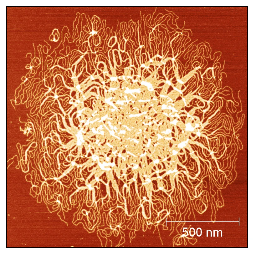
AFM image of metal complex–induced DNA condensation
3D In Vitro Models
Patient-derived organoids and 3D tumour models provide a physiologically relevant context for evaluating drug efficacy and selectivity beyond standard 2D cell culture. Our pipeline spans organoid establishment, drug treatment, and multimodal phenotypic readout.
Available
Under development
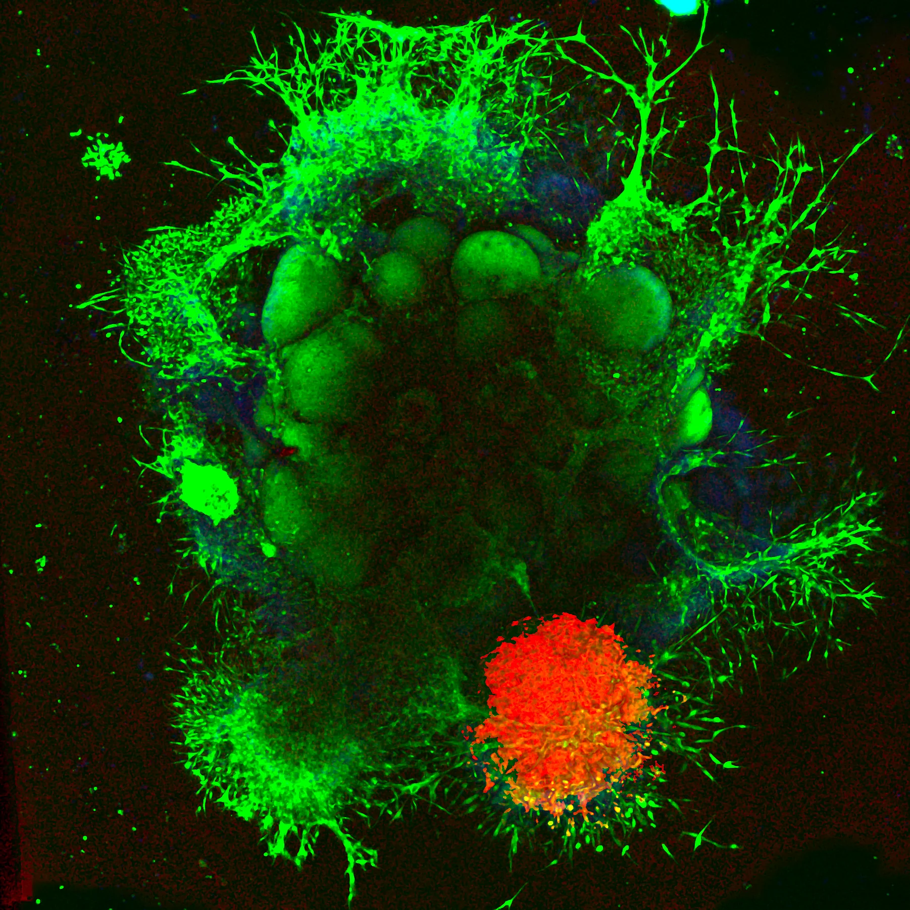
Glioblastoma organoid — bright-field microscopy
Blood-Brain Barrier Permeability
BBB penetration of candidate compounds is assessed using a validated model of human brain endothelial cells, providing a clinically relevant platform for CNS drug transport studies. Barrier integrity is confirmed before each experiment by transepithelial electrical resistance (TEER) measurements. Compound permeability is quantified by ICP-MS or fluorescence detection and validated against dextran tracers and FDA-approved reference drugs with established apparent permeability (Pₐₐₐ) coefficients.
BARRIER INTEGRITY
TEER measurement
PERMEABILITY QUANTIFICATION
ICP-MS and fluorescence
REFERENCE VALIDATION
Dextran tracers and FDA-approved drugs with known Papp
Cellular Pharmacology and Imaging
Comprehensive mechanistic profiling of anticancer candidates across panels of cancer and normal cell lines — and in 3D tumour spheroid models. We map intracellular drug distribution, reactive oxygen species generation, cell cycle perturbation, DNA damage, and programmed cell death pathways to build a full picture of drug action.
Analytical methods
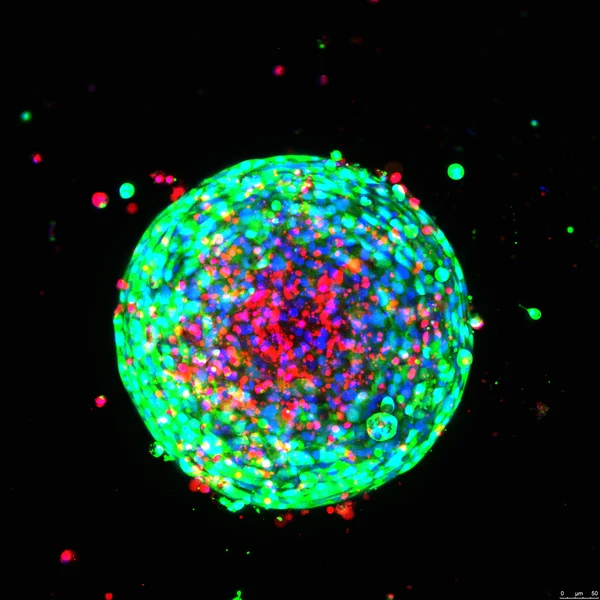
Non-small cell lung cancer spheroid after photodynamic therapy — calcein AM (metabolic activity, green), propidium iodide (dead cells, red), Hoechst (nuclei, blue)
Photodynamic Therapy
We investigate photoactivatable metal-based compounds — primarily iridium(III) and ruthenium(II) complexes — that generate cytotoxic reactive oxygen species upon light irradiation. Our programme spans mechanistic photochemistry, cellular phototoxicity profiling, and evaluation in advanced 3D tumour models including organoids and spheroids.
Hypoxic Conditions
PDT activity is assessed under normoxic and hypoxic atmospheres to model the oxygen-depleted tumour microenvironment — a critical factor for clinical translation of type I and type II photosensitisers.
Illumination Platforms
Variable-wavelength photoirradiation using a Luzchem photoreactor and dedicated well-plate illuminators, enabling systematic dose–response studies across multiple wavelengths in both 2D and 3D cell models.
Additional Focus Areas
Cancer Stem Cells and Therapy Resistance
Cancer stem cells (CSCs) represent a self-renewing subpopulation responsible for tumour initiation, recurrence, and resistance to conventional chemotherapy. We characterise the ability of novel metal-based compounds to target CSC-enriched populations in glioblastoma and colorectal cancer — addressing one of the key obstacles to durable remission.
Antimetastatic Therapy and Metastatic Processes
Metastasis accounts for the majority of cancer-related mortality, yet remains poorly addressed by standard cytotoxic agents. We evaluate the potential of metal-based compounds to interfere with key steps of the metastatic cascade — from epithelial-to-mesenchymal transition and cell migration to invasion and anchorage-independent growth — using mechanistic in vitro assays.
Alternative DNA and RNA Structures
Beyond canonical double-helical DNA, non-B-form structures such as G-quadruplexes, i-motifs, triplexes, and cruciform DNA play critical roles in gene regulation, telomere maintenance, and genome stability. We investigate how metal-based compounds interact with and stabilise these alternative architectures, opening new avenues for selective targeting of oncogene promoter regions and telomeric sequences.
Our Translational Philosophy
Every compound we characterize is evaluated against the backdrop of clinical relevance. By integrating fundamental biophysics with patient-derived organoid models, we bridge the gap between molecular mechanism and preclinical efficacy — accelerating the identification of next-generation anticancer agents.
Our TeamDepartment Members
A multidisciplinary team of biophysicists and cell biologists united by a common goal — advancing the science of metal-based cancer therapeutics.
Department Members
A multidisciplinary team of biophysicists and cell biologists united by a common goal — advancing the science of metal-based cancer therapeutics.

Vojtech Novohradsky, PhD.
Head of Department
Advanced in vitro models, translational oncology, patient-derived tumor models

Jaroslav Malina, PhD.
Deputy Head
In vitro pharmacology, interaction of compounds with alternative DNA/RNA structures, molecular interactions and thermodynamics
Hana Kostrhunova, PhD.
Senior Researcher
Cellular and molecular biology, pharmacology of experimental compounds, patient-derived CRC organoids, blood-brain barrier penetration studies

Lenka Markova, PhD.
Senior Researcher
Cellular and molecular biology, pharmacology of experimental compounds, CNS oncology, patient-derived glioblastoma organoids, cancer stem cells

Assoc. Prof. Olga Novakova
Senior Researcher
Biomacromolecular interactions, biochemistry, analytical and synthetic chemistry

Jakub Cervinka, PhD.
Postdoctoral Researcher
Cellular and molecular biology, pharmacology of experimental compounds, genomic and transcriptomic analysis, metabolomics
Prof. Jana Kasparkova
Senior Researcher
Cellular and molecular biology, biochemistry, DNA interactions
Prof. Viktor Brabec
Emeritus Professor
Founder of the department
PhD Students
Zuzana Nimmertondlova
PhD Student
Supervisor: Lenka Markova

Marie Svitelova
PhD Student
Supervisor: Vojtech Novohradsky
Sofia Sharkawy
PhD Student
Supervisor: Hana Kostrhunova
Technicians
Milada Korinkova
Technician
Team member details are placeholders — update with real names, roles, and photos before publishing.
Funding and ProjectsSupported Grants
Our research is funded through competitive national and European grants, reflecting the scientific merit and translational potential of our work in anticancer metallodrug discovery.
Supported Grants
Our research is funded through competitive national and European grants, reflecting the scientific merit and translational potential of our work in anticancer metallodrug discovery.
Induction of DNA damage in cancer cells by targeting alternative DNA structures by specific ligands. Relationship with their anticancer activity.
Czech Science Foundation
Biophysical analysis of metal-based drug-protein conjugates. Effect of carrier protein on transport of new organometallic antitumor compounds.
Institute of Biophysics CAS
Metal-based compounds as candidates for antimetastatic chemotherapy.
Czech Science Foundation
Medicinal biophysics and biochemistry of light-activated metallodrugs for targeted cancer therapy.
Czech Science Foundation
Selected WorksRecent Publications
A selection of peer-reviewed articles from the last five years of our research in inorganic medicinal chemistry, biophysics, and cancer biology.
Recent Publications
A selection of peer-reviewed articles from the last five years of our research in inorganic medicinal chemistry, biophysics, and cancer biology.
Chirality affects biological activity and interactions of azacryptands with alternative DNA structures
Malina J., Kostrhunova H., Cervinka J., Novakova O., Stoianova V., Müller C., Merten C.
Bioorganic Chemistry
Multitargeting Prodrugs that Release Oxaliplatin, Doxorubicin and Gemcitabine are Potent Inhibitors of Tumor Growth and Effective Inducers of Immunogenic Cell Death
Sarkar A., Novohradsky V., Maji M., Babu T., et al.
Angewandte Chemie International Edition
Ni(II) Cylinders Damage DNA in Cancer Cells and Preferentially Bind Y-Shaped DNA Three-Way Junctions Blocking DNA Synthesis
Malina J., Kostrhunova H., Brabec V.
Small
A novel benzothiazole-1,2,3-triazole-based arene osmium(ii) complex as an effective rhabdomyosarcoma cancer stem cell agent
Sharkawy S., Hernández-García A., Kostrhunova H., Bautista D., et al.
Inorganic Chemistry Frontiers
Photocatalytic arylterpyridine iridium(iii) complexes trigger oncosis in 2D and 3D cancer cell models via NADH oxidation
Romero-Castellón I., Markova L., Piernas-Muñoz M., Kostrhunova H., et al.
Inorganic Chemistry Frontiers
Photoactivatable Cyclometalated Ir(III) Compound Penetrates the Blood-Brain Barrier in 3D Spheroidal and Advanced 3D Organoid Models of Inherently Resistant and Aggressive Brain Tumors
Novohradsky V., Marco A., Svitelova M., Cutillas N., et al.
ACS Pharmacology & Translational Science
Excited-State Cis and Trans Pt(IV) Diamine Anticancer Complexes
Shi H., Kasparkova J., Ponte F., Kostrhunova H., et al.
Inorganic Chemistry
Antitumor platinum(II) complex 56MESS binds to DNA G-quadruplexes, downregulates expression of c-MYC and k-RAS oncogenes, and triggers DNA damage in cancer cells
Malina J., Kostrhunova H., Aldrich-Wright J., Brabec V.
Chemico-Biological Interactions
A Novel Substituted Benzo[g]quinoxaline-Based Cyclometalated Ru(II) Complex as a Biocompatible Membrane-Targeted PDT Colon Cancer Stem Cell Agent
Marco A., Kasparkova J., Bautista D., Kostrhunova H., et al.
Journal of Medicinal Chemistry
Antimetastatic activity of (arene)ruthenium(II) complex of 4-aryl-4H-naphthopyran
Pracharova J., Cyrikova T., Berecka M., Biersack B., et al.
Chemico-Biological Interactions
Dicobalt(ii) helices kill colon cancer cells via enantiomer-specific mechanisms; DNA damage or microtubule disruption
Song H., Kostrhunova H., Cervinka J., Macpherson J., et al.
Chemical Science
Multiaction Pt(IV) Prodrugs Releasing Cisplatin and Dasatinib Are Potent Anticancer and Anti-Invasive Agents Displaying Synergism between the Two Drugs
Markova L., Maji M., Kostrhunova H., Novohradsky V., et al.
Journal of Medicinal Chemistry
Interaction of dinuclear Co(III) cylinders with higher-order DNA structures
Malina J., Crowley J., Brabec V.
Chemico-Biological Interactions
New cyclometalated Ru(ii) polypyridyl photosensitizers trigger oncosis in cancer cells by inducing damage to cellular membranes
Cervinka J., Hernández-García A., Bautista D., Markova L., et al.
Inorganic Chemistry Frontiers
Open for CollaborationAdvancing Science Together
We welcome academic institutions, pharmaceutical companies, and biotech innovators to join forces with our department.
Advancing Science Together
We welcome academic institutions, pharmaceutical companies, and biotech innovators to join forces with our department.
Advancing Science Together
We welcome academic institutions, pharmaceutical companies, and biotech innovators to join forces with our department. Whether you seek access to our instrumentation, specialised expertise, or a full research partnership — we are ready to explore what we can achieve together.
CollaborationAnalytical Services
We offer access to our specialized instrumentation and expertise to academic partners, pharmaceutical companies, and biotech start-ups. Collaborative projects and service analyses are both welcome.
Analytical Services
We offer access to our specialized instrumentation and expertise to academic partners, pharmaceutical companies, and biotech start-ups. Collaborative projects and service analyses are both welcome.
Microscale Thermophoresis
Monolith Pico & Nano (NanoTemper)
Label-free and fluorescence-based measurement of molecular interactions directly in solution, without immobilisation. The Pico configuration reaches picomolar sensitivity for tight binders; the Nano variant uses intrinsic UV fluorescence for truly label-free analyses. Both formats are compatible with proteins, nucleic acids, small molecules, and nanoparticles.
- Binding affinity (Kd) from pM to mM range
- Protein–DNA and protein–small molecule interactions
- Label-free mode via intrinsic tryptophan fluorescence
- Low sample consumption (< 5 µL per measurement)
Nanoparticle Tracking Analysis
NanoSight NS300 (Malvern)
Visualises and tracks individual nanoparticles in suspension using light scattering and Brownian motion analysis. Delivers particle-by-particle size distributions, absolute concentration, and morphology data for liposomes, extracellular vesicles, protein aggregates, and metal-complex nanoformulations. Optional fluorescence mode enables selective tracking of labelled populations.
- Size distribution (10 – 2 000 nm)
- Absolute particle concentration (particles/mL)
- Fluorescence mode for labelled particle subpopulations
- EV/exosome characterisation and aggregation studies
Calorimetry
Nano DSC 6300 (TA Instruments) & VP-ITC (Malvern)
Complementary calorimetric techniques for full thermodynamic characterisation of biomolecular systems. DSC resolves thermal unfolding and stability of proteins and nucleic acids, revealing how drug binding affects structural integrity. ITC quantifies binding affinity, stoichiometry, enthalpy, and entropy of molecular interactions in a single label-free experiment.
- DSC: melting temperature (Tm), ΔHcal, cooperativity
- ITC: Kd, ΔH, ΔS, ΔG and stoichiometry
- Drug-induced stabilisation or destabilisation of targets
- No labelling or immobilisation required
Flow Cytometry
Amnis CellStream (Luminex)
High-throughput multi-parameter flow cytometer enabling simultaneous analysis of cell viability, apoptosis, cell cycle distribution, intracellular signalling, and surface marker expression. Its extended dynamic range and high sensitivity make it ideal for mechanistic pharmacology studies — from routine cytotoxicity profiling to in-depth cell death pathway dissection.
- Apoptosis (Annexin V/PI, caspase activation)
- Cell cycle analysis and γH2AX DNA damage quantification
- Mitochondrial membrane potential (JC-1, MitoTracker)
- ROS detection and intracellular metal staining
Circular Dichroism Spectroscopy
JASCO J-1500 (JASCO)
Polarised-light spectroscopy for probing the secondary and tertiary structure of proteins and nucleic acids, and for monitoring conformational changes induced by metal-based drug binding. The JASCO J-1500 covers a wide spectral range — from far-UV to visible — enabling simultaneous structural and electronic analysis. Temperature-controlled experiments allow real-time denaturation and melting studies.
- Protein secondary structure (α-helix, β-sheet, random coil)
- DNA/RNA conformational analysis (B-, A-, Z-form transitions)
- Drug-induced conformational changes in biomolecules
- Thermal denaturation and Tm determination
Interested in a collaboration?
Whether you need thermodynamic binding data, particle size characterization, or a full drug–target interaction study — we are open to academic and industrial collaborations. Reach out to discuss scope, pricing, and timelines.
Education and TrainingFrom hiPSC to Organoids
Intensive one-month hands-on training programmes taking participants from the fundamentals of human induced pluripotent stem cell culture through to advanced patient-derived tumour models — covering directed differentiation, tissue engineering, and pharmacological applications in two disease-focused tracks.
From hiPSC to Organoids
Intensive one-month hands-on training programmes taking participants from the fundamentals of human induced pluripotent stem cell culture through to advanced patient-derived tumour models — covering directed differentiation, tissue engineering, and pharmacological applications in two disease-focused tracks.
CNS Malignancies and Drug Discovery
From pluripotency to brain tumour model
A comprehensive one-month hands-on training programme bridging human pluripotent stem cell biology with neuro-oncology and preclinical drug evaluation. Participants master the full workflow from hiPSC maintenance through directed cerebellar differentiation, co-culture with patient-derived glioblastoma cells, and pharmacological assessment of candidate anticancer compounds — providing an advanced, clinically anchored research platform.
Who should attend
- Neuro-oncology researchers
- CNS drug discovery scientists
- PhD students and postdocs in brain tumour biology
Colorectal Cancer
From pluripotency to intestinal tumour model
An intensive one-month practical training programme focused on intestinal organoid biology and patient-derived colorectal cancer modelling. Participants acquire end-to-end competence — from hiPSC culture and intestinal directed differentiation to the derivation of patient organoids from clinical biopsy material, co-culture with tumour stroma, and high-content drug sensitivity profiling — delivering a translationally relevant platform for personalised oncology research.
Who should attend
- Colorectal cancer researchers
- Gastrointestinal oncology scientists
- Clinicians pursuing personalised medicine approaches
hiPSC Culture Fundamentals
Principles of human induced pluripotent stem cell biology; feeder-free maintenance and quality control; routine passaging, feeding schedules and mycoplasma testing; pluripotency authentication by immunofluorescence and flow cytometry.
Directed Differentiation
Step-by-step morphogen-guided protocols for cerebellar (Workshop A) or intestinal/colonic (Workshop B) lineage specification; optimisation of small-molecule and growth factor regimes; monitoring differentiation efficiency by qPCR and immunostaining.
Cryopreservation and Biobanking
Optimised slow-cooling and vitrification protocols for hiPSCs and organoids; controlled-rate freezing; thawing, recovery assessment and viability quantification; long-term storage strategy and biobank record management.
Patient-Derived Organoids
Derivation of organoids from primary patient tissue — surgical resections and diagnostic biopsies; tissue dissociation, single-cell isolation and ECM embedding; quality control, STR authentication and culture adaptation; ethical and regulatory considerations for clinical material.
Implantation and Tissue Modelling
Matrigel and synthetic hydrogel scaffold embedding; reconstruction of healthy and malignant tissue architectures in co-culture; morphological and functional assessment by brightfield, confocal and live-cell imaging; strategies for drug treatment and phenotypic readout in 3D.
Register Your Interest
Workshops are organised on demand and tailored to the participants' research background. Group placements and institutional partnerships are welcome. Contact us to discuss scheduling, capacity, and prerequisites.